Life
5 Things Radiologists See On Ultrasounds That Look Like Fuzz To You
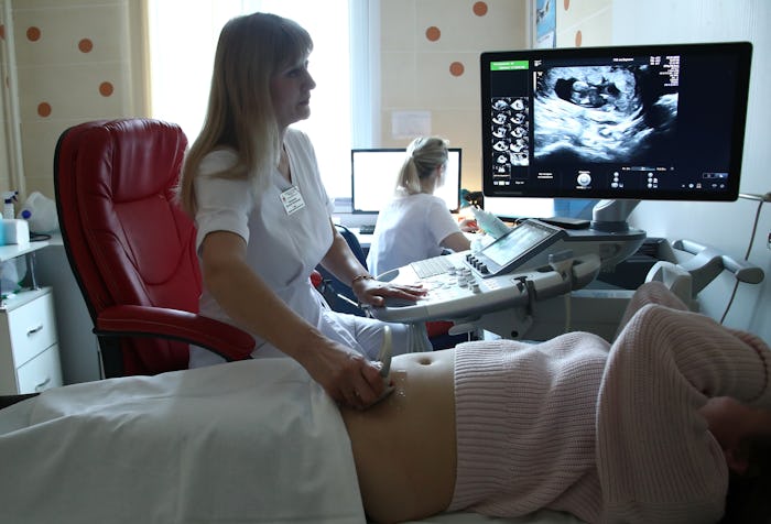
It’s exciting and sometimes emotional to see the first sonogram images of your baby, with their little head, tiny fists, and sweetly curled up body. But it can also feel more like watching a TV channel with a lot of static than looking at your own child for the first time, which is why it's so surprising when you realize all the things radiologists can see on fetal ultrasounds. While you're desperately trying to make out the shape of a foot or a hand in all the fuzz, they're spotting body parts (among other things) left and right. How do they do it?
It's important to talk about why radiologists are peering into your uterus in the first place. In addition to checking the amniotic fluid and the position of the baby, early ultrasounds are an accurate way to discern how far along the pregnancy is (much more so than, say, trying to recall when your last period was). This is known as "fetal biometry" and it measures the size of the fetus to determine your due date.
And if you've heard that ultrasounds are harmful to your baby? You can rest assured that's not the case. "There’s no truth to the idea that ultrasounds are stressful to the fetus," Sharyn Lewin M.D. tells Romper. "Ultrasounds are actually very safe in pregnancy; there’s really no detriment. In fact, ultrasound can be lifesaving because we can detect anatomic abnormalities or genetic syndromes that we wouldn’t know about otherwise. Ultrasounds are critical to providing the best prenatal care."
Read on for five things your gynecologist or radiologist can see during your ultrasounds that might not be so crystal clear to you, and maybe at your next appointment you can ask them to specifically point out some of the less obvious body parts (like teeny fingernails) yourself.
1The Baby Swallowing Amniotic Fluid

A lot of attention is paid to amniotic fluid on ultrasounds, especially how much or how little of it there is. If your doctor catches it at the right time, it's entirely possible to glimpse your baby taking a big gulp of amniotic fluid.
"Amniotic fluid is a sterile solution that surrounds and cushions the fetus inside the uterus during pregnancy. The fluid comes from the baby’s kidneys — it's fetal urine — and is absorbed when the fetus swallows it. The amount of fluid increases until the 36th week of pregnancy; after that, it slowly decreases," according to the Milton S. Hershey Medical Center website.
2A Full Head Of Hair
You may have heard that if you have heartburn during pregnancy, your baby is going to have a lot of hair. I know it sounds like on old wives' tale, but researchers at Johns Hopkins Medical Institutions actually found that there's at least some truth to this idea. One way you can know for sure that your baby is going to be born with some luscious locks? "Sometimes you can see hair on the ultrasound," Lewin tells Romper.
However, if no hair is detected, that doesn't necessarily mean you're having a bald babe. “Babies grow in amniotic fluid in the uterus, which means that their hair is wet,” Risa Klein, CNM, a midwife, told Romper in a previous article. “That can skew the image of hair length on an ultrasound.”
3The Upper Lip
As a person born (more than three decades ago) with a cleft lip, I was really surprised to hear that ultrasounds can detect the mouth and especially the upper lip. "Usually after 8-10 weeks you can see the upper lip," Lewin tells Romper. Later on, the palette and the jaw will be visible, too.
4 Gallstones Or Cysts In The Mother
Though baby is kind of the star of the show, your doctor should also be examining the mother during an ultrasound.
“We also want to look at the mom’s anatomy [including] the uterus, ovaries and fallopian tubes to make sure her anatomy looks normal, too," Lewin says. She adds that sometimes they spot gallstones or a mass on the ovary, which can be surprising if you’re not expecting it, but isn't usually a big deal. And if you've ever wondered why you have to go into ultrasounds with an uncomfortably full bladder?
“Ultrasounds are like sound waves of frequency greater than what the human ear can appreciate," Lewin tells Romper. "If an abdominal ultrasound is done, women often need to have their bladders full so that you can see better just based on how the frequency of the waves bounce off the bladder."
5The Beating Heart
In addition to the brain, stomach, kidneys, bladder, spinal cord, and abdominal wall (just to name a few) your doctor will be able to see different chambers of the heart, and cardiac activity.
“You can see the heart moving and beating," Lewin tells Romper. "It’s very cool. It’s reassuring for women to see the heart beating, to see the growth.”
Cue happy tears.
6Nuchal Thickness & Facial Structure
“You'll have something called a specialized or comprehensive exam [which] requires more specialized training by the [ultrasound tech]," Lewin says. "It looks for a more detailed exam of the anatomy than the standard fetal survey and so that's when we look for sex, a more detailed evaluation of the brain, the neck including the nuchal fold (or the nuchal thickness). There are some associations with down syndrome and other genetic syndromes based on the thickness of the fold or the skin."
In many cases your doctor will be able to see the fetus's face at this stage too (lips, nose, jaw, and ears) so trust them when they say your little one is cute.
7 7. Finger & Toe Nails

Believe it or not, your doctor or the ultrasound tech will be able to see tiny finger and toe nails. "Twelve weeks into your pregnancy, or 10 weeks after conception, your baby is sprouting fingernails," according to the Mayo Clinic. This explains why infants can be born with long fingernails and you will likely have to file or cut your baby's nails not long after birth. This can be a scary prospect, but it usually turns out just fine and can prevent the baby from scratching their face (or yours!).
Expert:
Dr. Sharyn Lewin, M.D., FACS founder and president of The Lewin Fund
This article was originally published on
